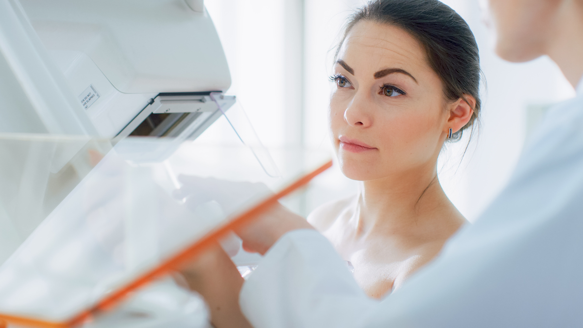
Misinformation surrounding mammograms can lead to anxiety. Get the facts about this important part of preventive care.
It’s not uncommon for women in their late 30s to consider what their first mammogram will be like. If you’re a woman over 40, you’ve probably had at least one, with another coming due within a year. And if you’re a man or a younger woman who’s experienced possible breast cancer symptoms, such as a lump, nipple discharge or dimpling of the skin, you may need one as well.
There are a lot of anxiety-inducing myths about mammograms, and women who’ve had bad mammography experiences can sometimes fear going back. Here are 5 facts to help clear things up so you can schedule your mammogram appointment with confidence.
1. Compression is needed but shouldn’t be very painful.
“Compression is an essential part of the mammogram,” explains Linda Larsen, MD, chief of breast imaging and director of radiology research in women’s health at USC Norris Comprehensive Cancer Center at Keck Medicine of USC.
In fact, inadequate compression can play a big role in poor image quality.
While it can be unpleasant, compression holds the breast in place and separates overlapping tissue to provide a clearer image so that small, hidden masses can be detected. It also reduces the amount of radiation that the patient is exposed to during the process.
All that said, communication and cooperation between the patient and technologist can help achieve compression with minimal discomfort.
2. The discomfort is brief and often manageable.
A common question doctors receive is, “Does a mammogram hurt?” The consensus among some patients is that they’re uncomfortable — but only briefly. Of course, breast sensitivity can vary according to different factors, such as hormone fluctuations. If you’re worried about a painful mammogram, talk to your technologist in advance. They can ease your worries and may have tips to help you prepare and minimize discomfort.
3. There are two types of mammograms: 2D and 3D.
In 2011, 3D mammography, also known as digital breast tomosynthesis, was approved by the U.S. Food and Drug Administration. Unlike conventional standard 2D mammography, which takes a single image of each breast, 3D mammograms move in an arc around the breast, capturing many more images from different angles in less time. This technique is more effective for a dense breast (with more glandular tissue than fatty tissue) and decreases the number of patients who need to come back for additional procedures. It’s also exceptionally accurate.
“Several research studies have shown that with 3D mammography, we can detect up to 40% more cancers than with 2D mammography alone,” says Larsen, who is also a professor of clinical radiology at the Keck School of Medicine of USC.
4. It’s recommended to start mammograms at age 40.
While there has been some debate on the appropriate age for a mammogram, the American College of Radiology recommends that women start getting yearly mammograms once they turn 40. If you have certain risk factors, your doctor may want you to start earlier.
“Women who are at a higher risk for breast cancer include those with genetic mutations or a strong family history of breast cancer, especially in a mother, sister or daughter,” Larsen says.
Regardless of age, anyone experiencing a lump or other symptoms should get a mammogram and possibly an ultrasound right away.
“Other signs of breast cancer may include thickening or swelling of the breast, change in size or shape of the breast, nipple discharge, redness or flakiness around the nipple, dimpling of the breast skin and a lump under the armpit,” Larsen says.
5. Mammograms save lives.
Mammography is the best tool available to screen for breast cancer. It can catch tumors before they can be felt in a monthly self-check, making treatment much more likely to be effective, Larsen explains. For example, in early cases where cancer cells are found only in the milk ducts, the likelihood of being cured is extremely high.
If cancer is found, your doctors will need to test the cells for certain proteins and genes to determine the best way to treat it. And as with many other cancers, it’s crucial to start treatment before it has a chance to spread.
Taking all of these factors into consideration, it’s clear that the expertise of the radiologist is critical. When choosing a provider, take a look at their reviews and success rates, and find out if they offer 3D mammography.
Topics
